There are several forms of macular degeneration, but the fastest growing form is age-related macular degeneration (AMD). AMD is the number-one cause of severe vision loss and legal blindness in adults over 60 in the U.S. As our population ages, and the “baby boomers” advance into their 60s and 70s, we will see a virtual epidemic of AMD.
Although it never causes total blindness by itself, age-related macular degeneration robs those affected of their sharp central vision and can dim contrast sensitivity and color perception. It destroys the clear, “straight ahead” central vision necessary for reading, driving, identifying faces, watching television, doing fine detailed work, safely navigating stairs and performing other daily tasks we take for granted.
The impact of developing AMD can be devastating to those who were independent and active prior to the onset of this impairment. Their visual world gradually diminishes into a vague blur, making ordinary daily activities challenging.

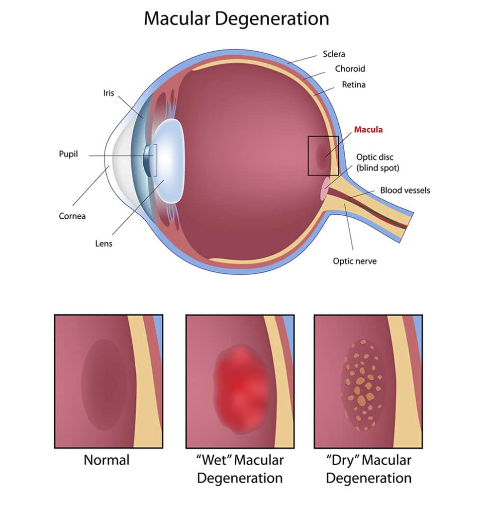
There are two types of AMD – “wet” or neovascular and “dry” or atrophic. There is no cure for AMD, but new treatments are available for the wet form of the disease. There is no treatment for the dry form, but vision rehabilitation and assistive devices can help people use their remaining vision effectively. If peripheral vision is not affected, it may be possible to see “out of the corner of your eye”.
When you go to your ophthalmologist for a regular check-up, you should receive a dilated retinal examination. If there are signs of macular degeneration, your doctor will notice the presence of drusen or new blood vessels beginning to grow in the macula.
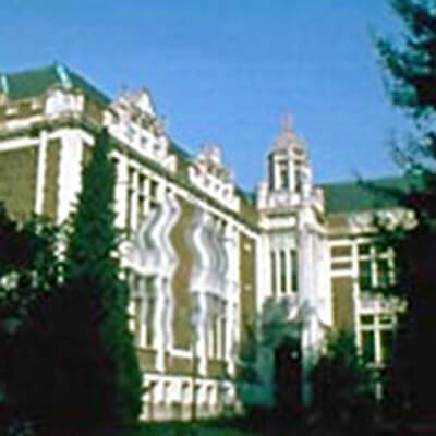
For many people, the first signs of AMD are things they notice themselves. For example, straight lines like doorways or telephone wires appear wavy or disconnected to them. When they look at a person, their face may be blurred while the rest of them is in focus. Lines of print may be blurred in the center or the lines may be crooked. For some people, there is a sudden blurring or loss of sight in the center of vision.
If you notice any distorted or missing areas of vision, consult your ophthalmologist promptly.

A new technology recently introduced promises to be an effective tool for early detection of AMD. Called the PreView PHP™ (Preferential Hyperacuity Perimetry), it identifies deviations in the retina using a special vision test.
A recent study showed that PHP testing yielded high percentages (over 80%) of sensitivity in detecting ‘wet’ AMD changes in 122 patients with intermediate-through-advanced AMD. This is a substantial improvement over previous methods of early diagnosis.
This exciting new technology is available at Clemson Eye, where we are committed to bringing the most advanced techniques and technology to care for your visual health.
To diagnose macular degeneration, your doctor may perform one or several tests. These are all non-invasive and painless. For two of them, you will have a dye injected into your arm with a very small needle. Most people experience very little discomfort, though the light from the camera can be glaring.
A technician will perform a test called a fluorescein angiogram. A dye is injected into the patient’s arm. The dye travels quickly through the body. When it reaches the back of the eye, a rapid sequence of photos of the retina is taken. The eye is dilated, so the retina can be seen through the wide open pupil. These photos show what changes have taken place in the retina and where abnormal blood vessels are located.
A procedure similar to fluorescein angiography, ICG angiography uses Indocyanine green dye, which can show more detail than flourescein angiography. The test uses a dye that is injected into the eye and then digital photographs are taken.
Your doctor may also take specialized photos that show the layers of the retina in cross section. There is no dye or injection involved. This special photograph lets the doctor see the thickness of the retina and any inflammation and fluids. As treatment continues, additional OCT images can show the effect of the therapy and can guide future treatment.
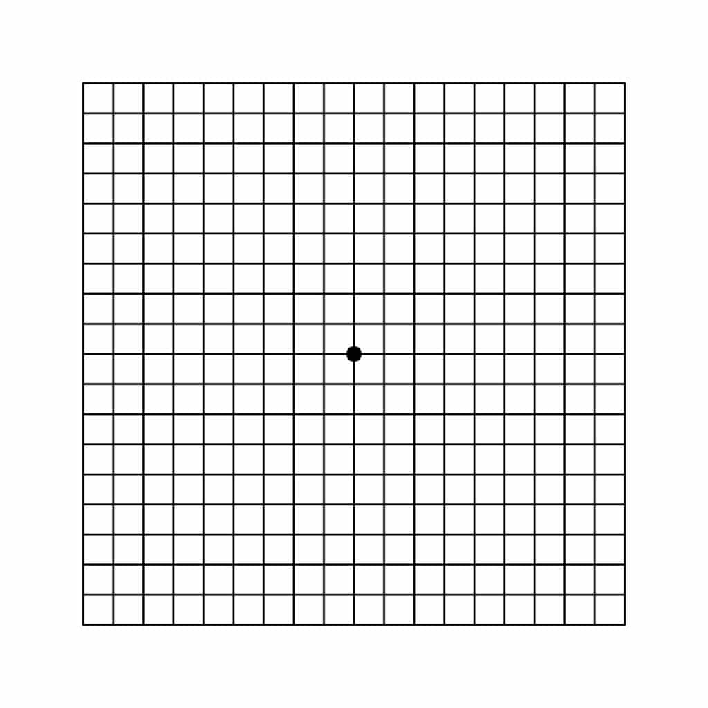
The Amsler Grid is a simple test of your central vision. It will alert you to any early changes that may indicate a problem with macular degeneration or a worsening of your condition.
The Amsler Grid looks like graph paper, with dark lines forming a square grid. Some versions have white lines on a dark background.
One of the first signs of macular degeneration can be wavy, broken or distorted lines OR a blurred or missing area of vision. The Amsler Grid can help you spot these early. Early detection of wet AMD is critical because laser or other treatments are most successful when performed before damage occurs. Since dry AMD can lead to wet AMD, the earlier the diagnosis, the better your chances of effective vision-saving treatment.
Your Clemson Eye ophthalmologist can tell you how often to use the Amsler Grid test.
All of the lines should be straight and the squares of a uniform size.
If you note any changes in the appearance of the grid, such as distortion, blurring, discoloration, dark or missing areas of the grid, or any other changes, see your eye doctor immediately. Do not wait to see if the changes will clear on their own. Timely treatment is vital to safeguarding your vision.

