Over 50% of the people who have glaucoma don’t know it since symptoms of vision loss, at least in the most common type of glaucoma, are generally not recognized until vision is lost.
Glaucoma affects about 2% of adults over the age of 40. Rarely, glaucoma can be present at birth or in childhood.
Glaucoma damages vision by gradually destroying the fine nerve fibers that travel from the retina and converge to become the optic nerve. The optic nerve connects your eye to your brain and carries visual information to your brain for processing.
Each nerve is like an electric cable containing about 1 million nerve fibers. As these nerve fibers are damaged and lost, you start to lose your peripheral or “side” vision. If the glaucoma remains untreated, the vision loss creeps in toward the center, first causing tunnel vision, and eventually blindness.
The cause of optic nerve damage in glaucoma is not known. Most patients with glaucoma have high fluid pressure, known as IOP, in the eye. Because of this, it is believed that fluid pressure plays a role in damaging the nerve.
The front chambers of the eye are filled with clear fluid called aqueous that is produced in one area called the ciliary body and drains out in another area called the angle. In glaucoma, the flow through the drainage structure of the eye becomes impaired, and fluid coming into the eye cannot get out, raising the intraocular pressure.
Many glaucoma patients have what is considered to be normal IOP during the day but it spikes up during the night. That means normal exams performed during the day may not identify the problem. In others the IOP may never reach abnormally high levels and they are considered to have Normal Pressure Glaucoma.
To make matters more complicated, not everyone with high IOP has glaucoma. They have what is known as ocular hypertension, a risk factor for developing glaucoma.
Although glaucoma is a treatable, but incurable disease that can cause blindness, this need not happen. If detected in time, it can be treated and precious vision can be preserved. Because glaucoma is generally without symptoms until it is too late and some vision is already lost, it is important for everyone to have regular eye exams.

Many people with glaucoma are unaware they have it until they have significant – and irreversible – vision loss. The cause of glaucoma is unknown, but there are several risk factors that increase the chances of developing glaucoma.
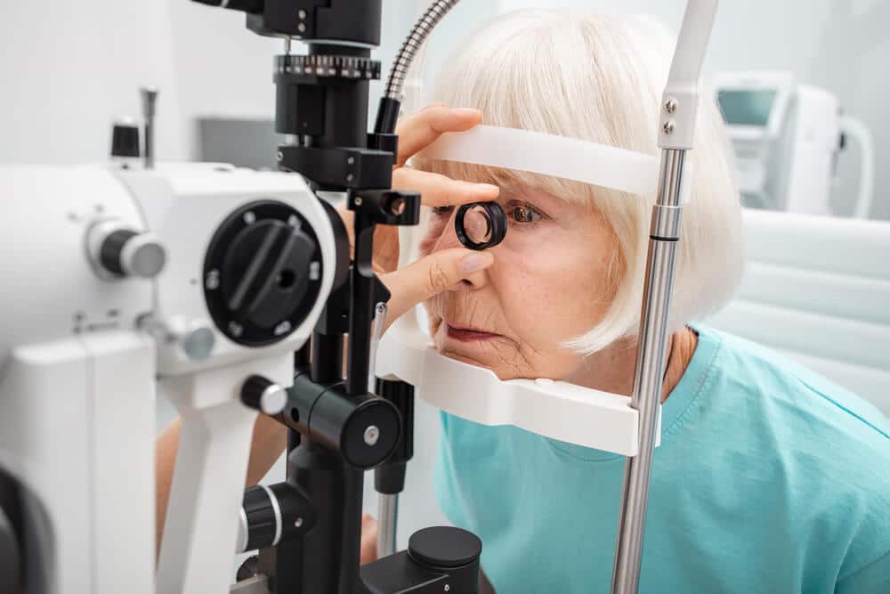
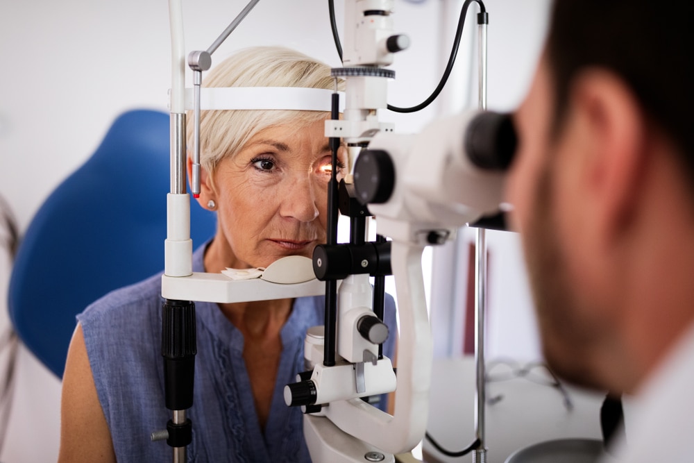
Individuals at risk for glaucoma and everyone over the age of 40 should have regular, comprehensive eye exams that include careful evaluation of the optic nerve and measurement of eye pressure.
Most glaucoma patients are seen by their eye doctor two to four times a year. During these exams, the IOP is measured, and the inner structure of the eye, including the retina, optic nerve head and optic disc, is checked. Maintaining pressure at the normal level is a primary goal of treatment.
Other assessments will be necessary at least once a year to give you the best opportunity to avoid further loss of sight. For example, a dilated examination is performed to evaluate the optic nerve. Periodically, it is also necessary to check the angle of the eye in a test called Gonioscopy. This is done to monitor the fluid drainage, which is important in maintaining proper pressure.
Each year, a Retinal Tomography test should be performed, which is a diagnostic test used to monitor the optic nerve head, as well as identify potential retinal disease. This is a very precise test that allows the doctor to identify early changes or damage to the optic nerve and the retinal nerve fiber.
Every several years, Fundus Photos are taken. These are photographs of the entire inner surface of your eyeball (fundus). These photos are vital in documenting any changes and determining if these changes are due to glaucoma or other related problems.
The Visual Field test is performed at least once a year. This extremely important test tracks the eye’s ability to correctly respond to light on all parts of the retina; areas that are damaged by glaucoma will show no response. Visual fields are necessary to determine if there is any damage to the optic nerve that may not be detected by simply looking at it. This test, which takes 30 minutes to an hour to perform, is essential to controlling your glaucoma.
If you are diagnosed with glaucoma, treatment is available to save your vision. Unfortunately, the vision that is already lost cannot be returned. That is why it is so important to have regular eye exams, particularly if you have any of the known risk factors for glaucoma.
The goal of glaucoma treatment is to lower IOP, stop the optic nerve damage and preserve your remaining vision. Several kinds of treatment are available to lower IOP. These include eye drops, laser therapy, and surgery.
Eye drop medications lower IOP by either reducing the amount of fluid entering the eye or increasing the amount of fluid exiting the eye. There are several different kinds of glaucoma medications, and each differs in terms of both its ability to lower IOP and its potential side effects.
We are dedicated to providing our patients the best care possible but you must do your part as well. It is imperative that you follow our instructions concerning medications and dosages, and that you commit to keeping your follow-up appointments.
During your visit please bring your medication(s) so we can ensure you are taking the correct medicine at the correct times. We also encourage you to write down any questions that you have so they can be answered and discussed.
Until recently, laser therapy was used primarily when medications failed to successfully lower IOP, or for patients who could not tolerate medications due to the side effects.
Recent advances in laser therapy have produced lasers so safe and effective that they are being used as first-line treatment instead of medications. If medications and / or laser therapy fail to bring the IOP down to a safe range, surgery can lower IOP.
The iStent is the smallest medical device ever approved by the FDA and is placed in your eye during cataract surgery. It is so small, you won’t be able to see or feel it after surgery but it will be continuously working to help reduce your eye pressure.
Implanting the iStent does not significantly lengthen the time a patient spends in surgery and has a safety profile comparable to cataract surgery alone. The iStent helps control eye pressure while reducing or eliminating the need for drops. This is a significant advantage, as it helps address the high rate of medication non-compliance among glaucoma patients.
As many as 90% of patients do not adhere to their prescribed regimen of drops and more than half stop using them completely. This is a serious problem as when pressure in the eye is out of control, it can increase the risk of permanent vision loss.
So far, iStent results have been very positive with about 68% of patients remaining medication free 12 months after their procedure. 2
iStent works like the stents used to prevent heart attacks and strokes. When blood vessels get clogged, a stent creates access to the vessel flow. While a highly innovative technology, how iStent works is elegantly simple:
If you have glaucoma, over time the eye’s natural drainage system becomes clogged. iStent creates a permanent opening through the blockage to improve the eye’s natural outflow. Restoring this mechanism lowers and controls pressure within the eye.
Dr. Joseph Parisi, Medical Director at Clemson Eye, has been trained in the iStent surgery and is one of a handful of surgeons in South Carolina approved to implant this device.
Implanting the iStent during cataract surgery is covered by most insurance plans, Medicare and Medicaid.
Selective Laser Trabeculoplasty (SLT) uses a laser to selectively target pigmented cells in the trabecular meshwork of the drainage angle. A biologic response is triggered leading to enhanced fluid outflow and reduced IOP.
This painless procedure is performed in the office in a matter of minutes. The laser is directed through a mirrored contact lens held against the eye. Effectiveness is evaluated 4-8 weeks later. SLT on its own is effective in controlling IOP over 90% of the time as a primary treatment.
When used as a second-line treatment in patients using glaucoma drops, many patients were able to discontinue one or more of their eye drops and maintain good IOP control.
The effect appears to last, with only 25% needing a repeat treatment within five years. Repeat treatments can produce similar effects, but the amount of IOP reduction is less with each successive re‑treatment.